








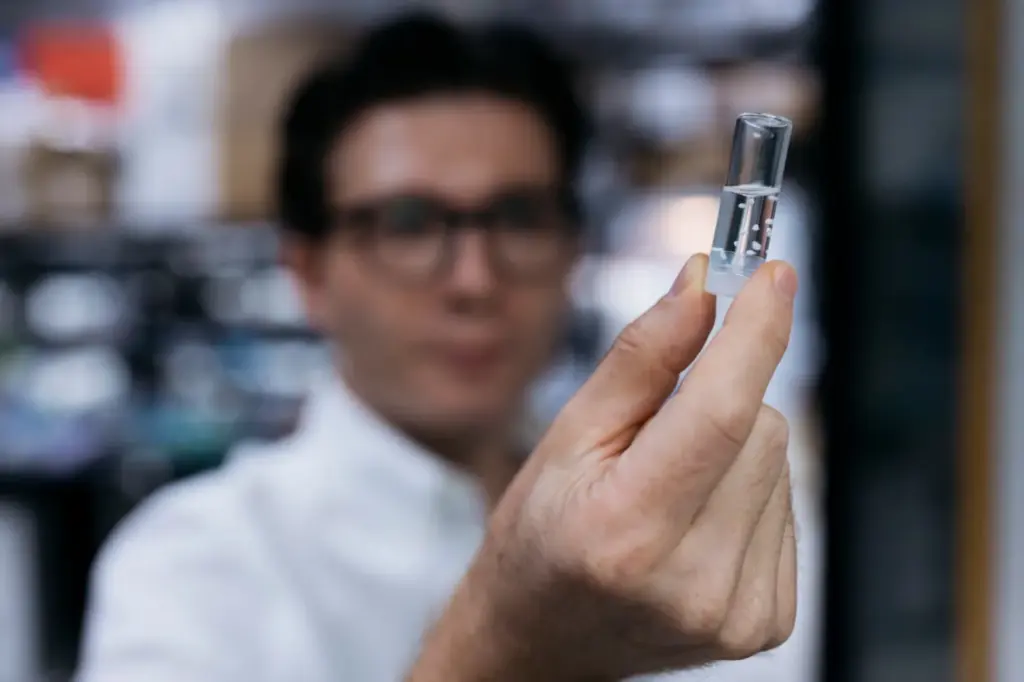
Neuroscientist Sergiu Pasca shows a transparent jar in which tiny white balls float. Each one is a cerebral cortex organoid, a millimetric replica of the brain area where complex thought, language, and the secret of the human mind reside. Pasca’s team has implanted these seeds into the brains of mice that had previously been modified so they would not develop a cerebral cortex. The experiment, unique in the world, has resulted in healthy animals whose brains are an integrated mix of human and mouse. They are the animals with the most human neurons grafted into their brains ever born, the scientist highlights in conversation with EL PAÍS.
Read more Trump decertifies Colombia again in the war on drugs
These animals allow for the first time to investigate “characteristics of the human brain that until now were inaccessible,” emphasizes Pasca, a physician and researcher at Stanford University in the United States. He refers to studying the most characteristic human neurons of our species at the molecular and cellular level in a living organism, not in a cadaver sample or a laboratory cell culture, as until now. The scientist, born in Romania 44 years ago, believes this advance paves the way to seek treatments against autism, schizophrenia, cerebral palsy, and dementia. In the future, Pasca points out, it will be possible to generate a mouse with a cerebral cortex made with the neurons of each patient.
The details of the experiment, described this Wednesday in Nature, seem like science fiction. It all begins with skin cells taken from healthy volunteers, to which scientists apply a cocktail of proteins to return them to their embryonic state, when they are capable of becoming any tissue in the body. Then they make them differentiate into the type of stem cells from which cerebral cortex neurons arise until composing organoids, those white balls already with a three-dimensional structure that Pasca shows during the interview, conducted by teleconference.
In previous experiments, the same team successfully implanted human neurons into the brains of rats to control their behavior with laser light, but their growth was limited by lack of space inside the skull — human neurons are much larger. The researcher had also demonstrated that organoids from different brain areas can be made and fused, as if they were pieces of a living puzzle.
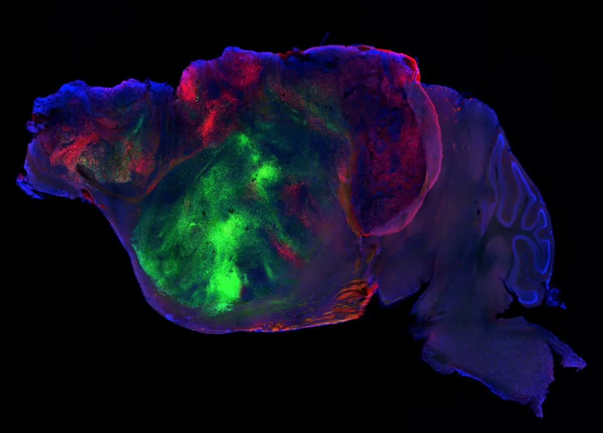
These spheres with hundreds of thousands of human neurons, grafted into mice two days after birth, begin to grow, vascularize, and connect with the rest of the mouse brain in which they reside, for example, sensory functions and movement. Since the rodent lacks its own cortex, the human neurons expand to compose more than 90% of the cortex and half of the entire brain of the mice. Action must be quick, as this window of “plasticity” to fuse closes about two weeks after implantation.
The cerebral cortex of these mice is comparable to that of a human fetus in the sixth month of gestation, explains Pasca, although with important differences. At that time, the human brain already has more than 16 billion neurons, while the modified mice have about four million of these cells in their cortex. A normal mouse brain, on the other hand, contains about 14 million neurons because they are much smaller. What is inside the brain of these modified animals is not exactly a human cerebral cortex, as some cell types are missing — and the real structure and size — but it is a very useful model for researching our “most inaccessible organ,” argues Pasca.
Cerebral palsy
In a proof of concept, the researchers cut off the oxygen flow to the brains of their humanized mice. Normal mice are very resistant to this aggression, while in humans, a small lack of air can trigger lifelong cerebral palsy. The lesions observed in animals with humanized brains resemble those seen in patients with cerebral palsy, which affects about 18 million people worldwide. The team suggests that their mice allow for the first time to study in vivo the impact of these currently irreversible lesions, and even to seek ways to cure them.
In 2024, Pasca’s team implanted human brain organoids made with cells from a child suffering from Timothy syndrome, a rare genetic disorder that causes severe autism and epileptic seizures, into rats. The work allowed scientists to design a genetic treatment that reversed the problem in the implanted rats. The team plans to begin testing that cure in humans in a clinical trial at the end of this year or early next year, explains Pasca.
Read more The composer Laura Vega and the performer Cecilia Bercovich, National Music Award winners 2026
Garikoitz Lerma-Usabiaga, an Ikerbasque researcher at the Basque Center on Cognition, Brain and Language (BCBL) in San Sebastián, participated in the study by characterizing the brain microstructure using diffusion magnetic resonance imaging. This technique measures the movement of water molecules to determine tissue organization and map the trajectory of neuronal pathways. “The technique finds that water molecules prefer to travel in the direction from the human organoid to the mouse brain, suggesting connectivity between the two,” explains the neuroscientist.
The researchers had another surprise: these mice generated a type of neuron that had never been isolated in living organisms. They are Von Economo neurons, a type of neural cell discovered in 1925 that is not abundant. At first, they were thought to be exclusively human, but later were found in primates and other mammals with large and complex brains such as elephants, whales, and dolphins. These neurons are located in areas of the cortex responsible for sociability and decision-making, and their premature death characterizes frontotemporal dementia, a rare variant that appears between 40 and 60 years of age. It is possible that these large neurons appeared in the humanized brains of the mice because, for the first time, there was enough space to grow. They may now allow testing drugs against the disease.

Although their cortex is made of human neurons, the behavior of these animals does not differ much from normal mice, assures Pasca. If anything, they are somewhat slower in memory and reflex tests because the connection between human and rodent brain “is not perfect.” This is because there are more than 70 million years of separate evolution between one species and the other.
Ethical issues
The development would be very different if these human organoids were implanted in monkeys, something that should not be done for ethical reasons, according to Pasca. Before conducting their experiments in mice, his team subjected them to supervision by an independent panel of experts in bioethics, lawyers, evolutionary biologists, patient associations, and philosophers, who approved the work. “It is a very delicate topic and it is easy for misunderstandings to arise if we do not convey the nuances well,” Pasca acknowledges. The researcher argues his refusal to replicate this experiment with apes: “In species closer to us, human neurons would have much more space to grow and the integration would be much better. I think we should be very cautious about this type of experiment and not carry them out unless there is a very clear justification.”
Biochemist Daniel Tornero, head of the stem cell and brain injury laboratory at the University of Barcelona, knows Pasca’s work well and calls it “incredible.” “Psychiatric diseases are the hardest to model because mice do not suffer from them, and their cerebral cortex is much smaller,” he explains. “This new work is interesting because it allows you to model human diseases with human cells,” he highlights.
In 2019, a Chinese team in which the Spaniard Juan Carlos Izpisua participated created the first monkey-human chimera: a macaque implanted with stem cells obtained from people. The goal was to explore the use of animals to grow human organs for transplants. The term chimera referred to the mythological animal that vomited flames and had the head of a lion, the belly of a goat, and the tail of a dragon. For Tornero, the animals created by Pasca are chimeras, although the Stanford researcher is careful to use that term, possibly to avoid controversy. Tornero reflects that it is difficult to know if there is anything human in the behavior or thought of these mice. “Human neurons send axons [projections] inside the mouse brain, but the data are not yet enough to know if those connections are superior,” he opines. It is a real possibility that was already demonstrated, on a much smaller scale, in 2013, when a team injected 300,000 human glial cells — responsible for supporting and immunity in the brain — into the brains of mice. The modified rodents had better memory and learned faster than normal ones.
For now, these new animal models only serve to study diseases that arise during embryonic development. Human neurons grow 20 times slower than those of mice. Even if one of these animals lived up to two years, its maximum life expectancy, and the equivalent of an elderly human, its cerebral cortex would still be like that of a two-year-old baby.